Pretty good description. Few extra points.
-you don’t need to don’t need to do PCR to have enough Nucleic acids to visualize on the gel. Often you use gels to visualize things like purified plasmids. You can also digest your plasmids with enzymes to confirm genomic insert.
-these type of gels have many uses outside of direct comparisons, for example you can use the gel purity DNA of a particular size by cutting out the band, melting the gel and using a silica column to pull the DNA out of the melted gel slice. This is super common when you do molecular cloning.
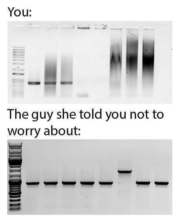
For what is possibly wrong with the top gel.
the wavy line in some bands indicates uneven current, this can be due to sample composition before loading the gel, like having cellular debris along with your DNA or a mismatch between the buffer used to make the gel and the running buffer.
-the blurred bands can be a lot of things. It might just be inherited to the sample type, for example raw genomic DNA will look like this because you’ll have enzyme that degrade DNA, and a lot of mid replication chromosomes resulting in fragments existing across a spectrum of sizes equally. Additionally the it could also be user error with overloaded well or not enough loading buffer in the sample so it’s not pushed in to gel evenly, or it was run to fast.
Overall the top gel just looks like crap and the bottom gel looks great, but depending on what was run you really don’t know what the gel should look like, if I was testing endonuclease activity the top gel would probably be what I was hoping to get. If was doing PCR on single gene, the bottom one is better.


Pretty good description. Few extra points. -you don’t need to don’t need to do PCR to have enough Nucleic acids to visualize on the gel. Often you use gels to visualize things like purified plasmids. You can also digest your plasmids with enzymes to confirm genomic insert. -these type of gels have many uses outside of direct comparisons, for example you can use the gel purity DNA of a particular size by cutting out the band, melting the gel and using a silica column to pull the DNA out of the melted gel slice. This is super common when you do molecular cloning.
For what is possibly wrong with the top gel.
-the blurred bands can be a lot of things. It might just be inherited to the sample type, for example raw genomic DNA will look like this because you’ll have enzyme that degrade DNA, and a lot of mid replication chromosomes resulting in fragments existing across a spectrum of sizes equally. Additionally the it could also be user error with overloaded well or not enough loading buffer in the sample so it’s not pushed in to gel evenly, or it was run to fast.
Overall the top gel just looks like crap and the bottom gel looks great, but depending on what was run you really don’t know what the gel should look like, if I was testing endonuclease activity the top gel would probably be what I was hoping to get. If was doing PCR on single gene, the bottom one is better.